Abstract
Rationale: Improving imaging-based response following neoadjuvant chemotherapy (NAC) in breast cancer assessment could obviate the need for histologic confirmation of pathological complete response (pCR) and facilitate de-escalation of chemotherapy or surgery. Fibroblast activation protein inhibitor (FAPI) PET/MRI is a promising novel molecular imaging agent for the tumor microenvironment with intense uptake in breast cancer. We assessed diagnostic performance of follow-up breast FAPI-PET/MRI in classifying response status in local breast cancer and lymph node metastases after completion of NAC and validated this approach immunohistochemically.
Methods: In women that completed NAC for invasive breast cancer, follow-up Gallium-68-FAPI-46 PET/MRI and corresponding FAP immunostainings were retrospectively analyzed. Metrics of FAPI uptake and FAP immunoreactivity in women with or without pCR were compared with the Mann-Whitney U test. Diagnostic performance to detect remnant invasive cancer was calculated for tracer uptake metrics using receiver operating characteristics curves and for blinded readers’ visual assessment categories of PET/MRI and MRI alone.
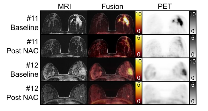
Results: 13 women (mean age 47 years ± 9) were evaluated. 7/13 women did achieve pCR in the breast and 6/10 pCR in the axilla. FAP immunoreactivity was significantly associated with response status. FAPI-PET/MRI mean breast tumor-to-background ratio (TBR) for pCR was 0.9 (range, 0.6-1.2) and for no pCR 2.1 (range, 1.4-3.1), P = .001. Integrated PET/MRI could classify breast response correctly in all 13 women based on readers’ visual assessment or TBR. Evaluation of MRI alone resulted in at least 2 false positives. For lymph nodes, PET/MRI readers classified at least one case falsely negative, whereas MRI alone resulted in 2 false negatives and 1 false positive.
Principal Conclusion: This is the first analysis of FAPI-PET/MRI for response assessment after NAC for breast cancer. The diagnostic performance of PET/MRI in a small study sample trended towards a gain over MRI alone, clearly supporting future prospective studies.


![FAP Expression in Renal Tumors Assessed by [68Ga]Ga-FAPI-46 PET Imaging and FAP Immunohistochemistry: A Case Series of Six Patients](https://sofie.com/wp-content/uploads/2025/12/info.ibamolecular-scaled-500x383.jpg)


![[68Ga]Ga-API-46 PET accuracy for cancer imaging with histopathology validation: a single-centre, single-arm, interventional, phase 2 trial](https://sofie.com/wp-content/uploads/2025/09/image-500x383.png)
