
Annika Hess, Alexandra Renko, Andreas Schäfer, Mira Jung, Daniela Fraccarollo, Jan D Schmitto, Johanna Diekmann, Thomas Thum, Frank M Bengel, Johann Bauersachs, James T Thackeray, Jochen Tillmanns
Abstract:
Purpose: Myocardial infarction (MI) triggers complex cellular responses essential for tissue repair and remodeling, including myofibroblast activation. Fibroblast activation protein alpha (FAP) identifies activated myofibroblasts post-MI, however its spatial distribution relative to the scar and area at risk (AAR) is unclear. Non-invasive FAP-imaging with PET radiotracer 68 Ga-FAPI-46 shows uptake beyond the infarct scar. We therefore aimed to characterize FAP expression in the AAR using a myocardial ischemia-reperfusion (MI/R) model in mice.
Procedures: We induced MI/R in male C57BL/6N mice. The AAR was identified by in vivo lectin staining, and expression of FAP, CD68, and hypoxic tissues were measured using immunohistochemistry. Spatial FAP was further interrogated by 68 Ga-FAPI-46 in mice by autoradiography and humans by PET. Additionally, human cardiac tissues from acute MI patients were examined for fibroblasts and inflammatory cells by expression of FAP, CD13, and α-smooth muscle actin.
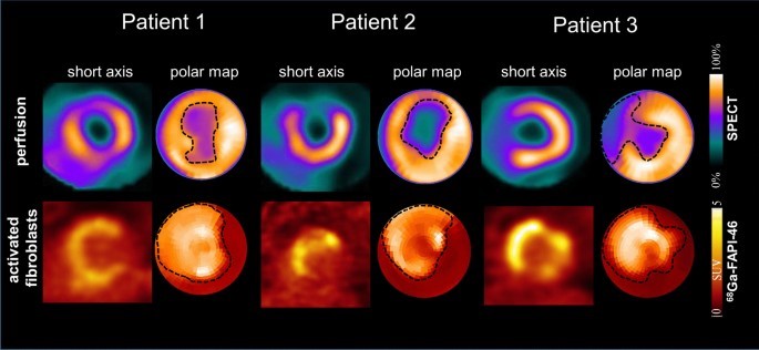
Results: FAP expression peaked three days post-MI/R predominantly within the AAR (p < 0.05 vs. d0). Consistent between murine models and human tissues, FAP+ myofibroblasts accumulated within the infarct scar and borderzone, occasionally extending into non-ischemic myocardium. CD68+ macrophages peaked similarly at three days post-MI/R (p < 0.05 vs. d0). FAP expression weakly correlated with CD68 but not with extent of ischemic or hypoxic territory post-MI/R. FAP imaging in mice and humans revealed aligned non-uniform 68 Ga-FAPI-46 uptake extending from the infarct scar into surviving myocardium after MI.
Conclusions: Our findings demonstrate a distinct FAP expression pattern post-MI/R. The alignment of ex vivo 68 Ga-FAPI-46 signal with myofibroblasts in the AAR supports its identification of a unique substrate in myocardial injury complementing other non-invasive imaging measurements of perfusion, viability and fibrosis.
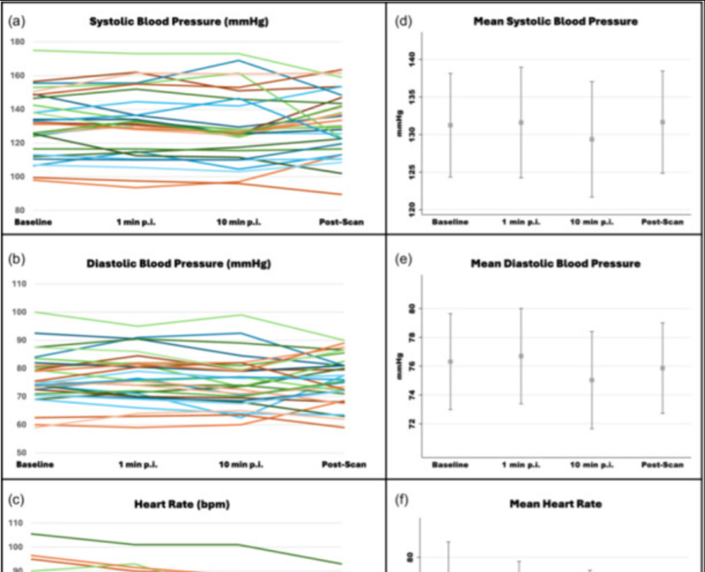
![Safety of [68Ga]Ga-FAPI-46 PET/CT including hemodynamic measurements and patient-reported outcomes in cancer patients](https://sofie.com/wp-content/uploads/2026/05/NewImage-500x383.jpg)
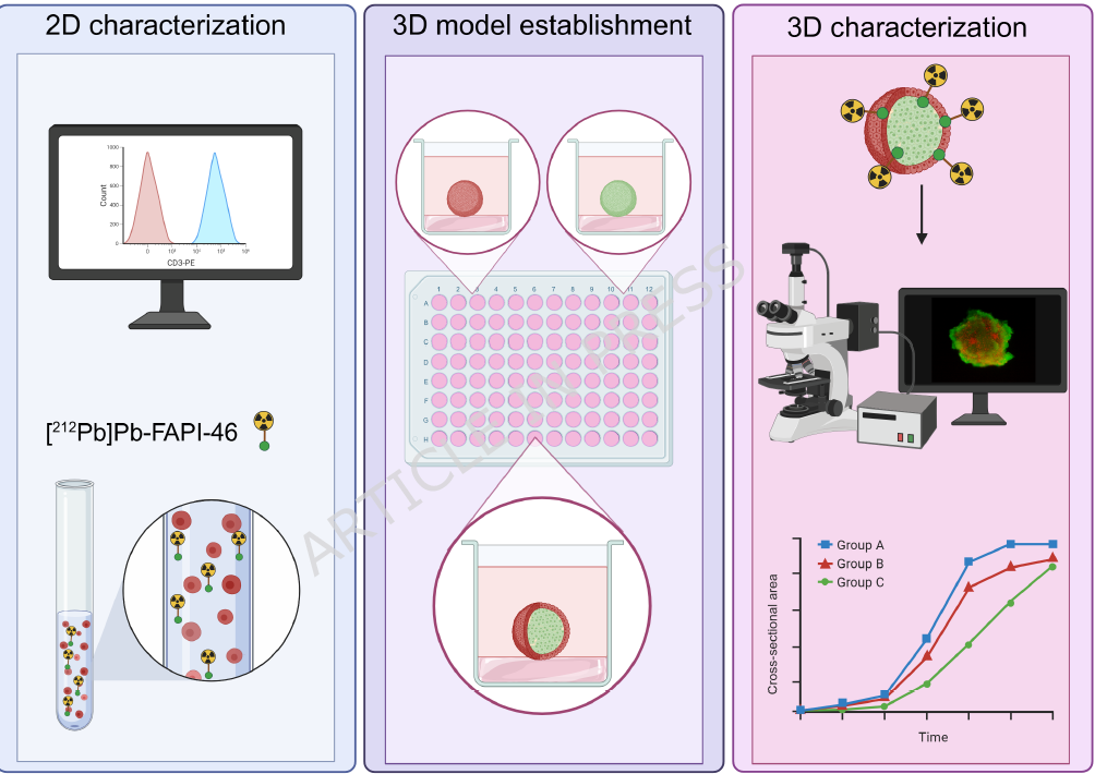
![A 3D co-culture model with fibroblast-restricted FAP expression for cancer therapy using [212Pb]Pb-FAPI-46](https://sofie.com/wp-content/uploads/2026/06/A-3D-co-culture-model-with-fibroblast-500x383.png)