
Lukas Kessler, Nader Hirmas, Kim M. Pabst, Rainer Hamacher, Justin Ferdinandus, Benedikt M. Schaarschmidt, Aleksandar Milosevic, Michael Nader, Lale Umutlu, Waldemar Uhl, Anke Reinacher-Schick, Celine Lugnier, David Witte, Marco Niedergethmann, Ken Herrmann, Wolfgang P. Fendler and Jens T. Siveke
Abstract
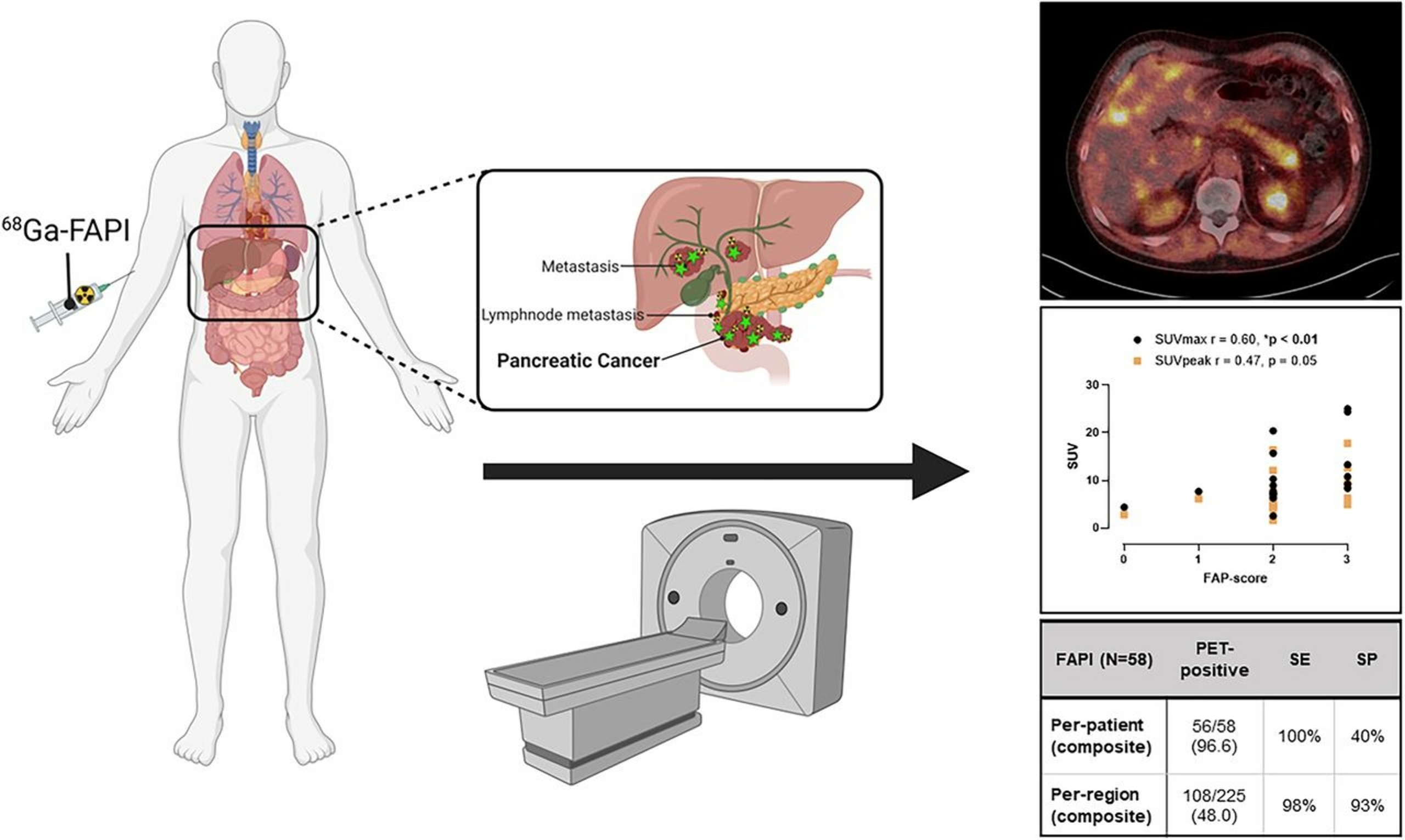
The fibroblast activation protein (FAP) is highly expressed on carcinoma-associated fibroblasts in the stroma of pancreatic cancer and thus is a promising target for imaging and therapy. Preliminary data on PET imaging with radiolabeled FAP inhibitors (FAPIs) demonstrate superior tumor detection. Here we assess the accuracy of FAP-directed PET in patients with pancreatic cancer.
Methods:
Of 64 patients with suspected or proven pancreatic cancer, 62 (97%) were included in the data analysis of the 68Ga-FAPI PET observational trial (NCT04571086). All of these patients underwent contrast-enhanced CT, and 38 patients additionally underwent 18F-FDG PET. The primary study endpoint was the association of 68Ga-FAPI PET uptake intensity and histopathologic FAP expression. Secondary endpoints were detection rate, diagnostic performance, interreader reproducibility, and change in management. Datasets were interpreted by 2 masked readers.
Results:
The primary endpoint was met: The association between 68Ga-FAPI SUVmax and histopathologic FAP expression was significant (Spearman r, 0.48; P = 0.04). For histopathology-validated lesions, 68Ga-FAPI PET showed high sensitivity and positive predictive values (PPVs) on per-patient (sensitivity, 100%; PPV, 96.3%) and per-region (sensitivity, 100%; PPV, 97.0%) bases. In a head-to-head comparison versus 18F-FDG or contrast-enhanced CT, 68Ga-FAPI detected more tumor on a per-lesion (84.7% vs. 46.5% vs. 52.9%), per-patient (97.4% vs. 73.7% vs. 92.1%), or per-region (32.6% vs. 18.8% vs. 23.7%) basis, respectively. 68Ga-FAPI PET readers showed substantial overall agreement on the basis of the Fleiss κ: primary κ, 0.77 (range, 0.66–0.88). Minor and major changes in clinical management occurred in 5 patients (8.4%) after 68Ga-FAPI PET.
Conclusion:
We confirmed an association of 68Ga-FAPI PET SUVmax and histopathologic FAP expression in pancreatic cancer patients. Additionally, we found high detection rate and diagnostic accuracy, superior to those of 18F-FDG PET/CT. 68Ga-FAPI might become a powerful diagnostic tool for pancreatic cancer work-up.
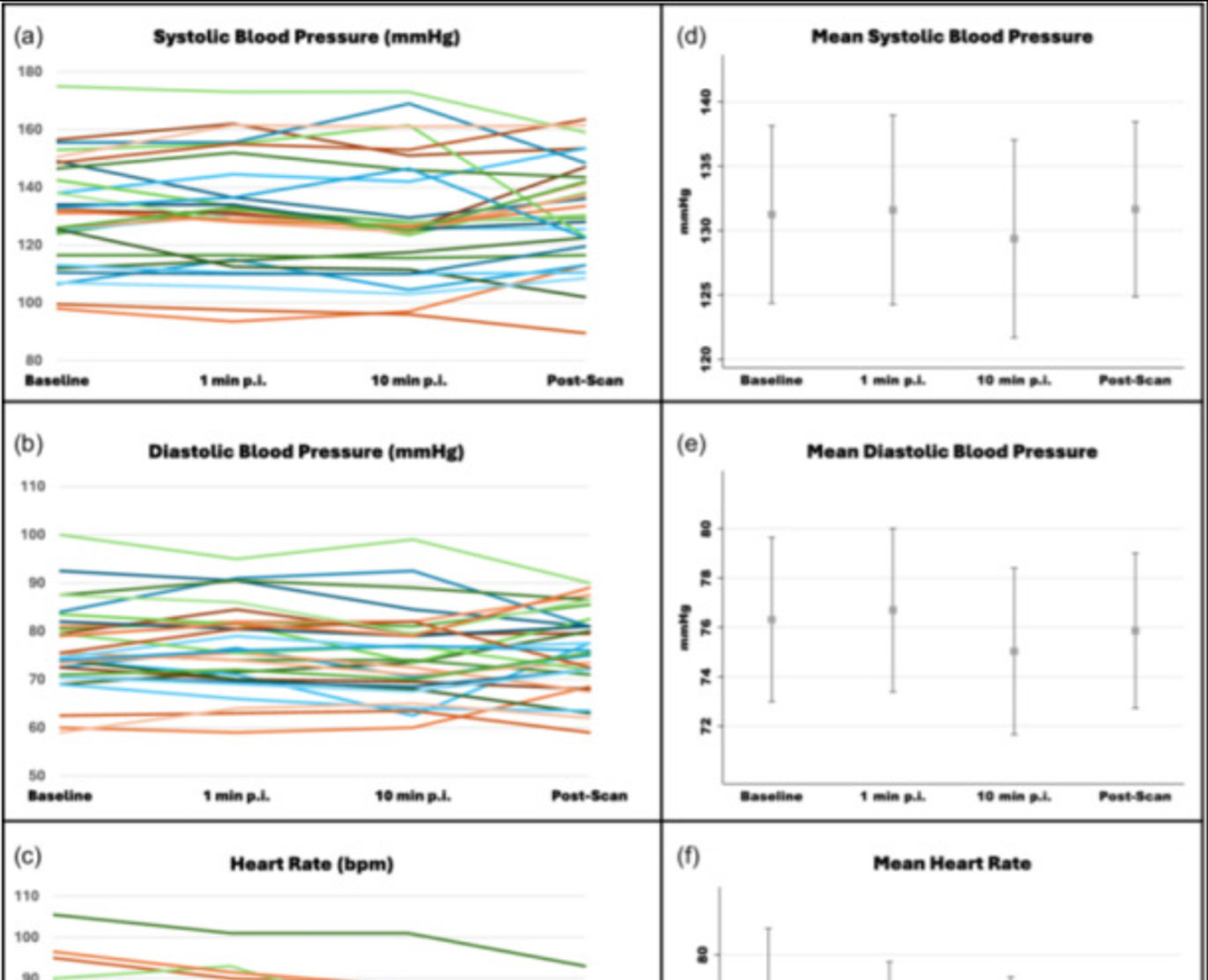
![Safety of [68Ga]Ga-FAPI-46 PET/CT including hemodynamic measurements and patient-reported outcomes in cancer patients](https://sofie.com/wp-content/uploads/2026/05/NewImage-500x383.jpg)
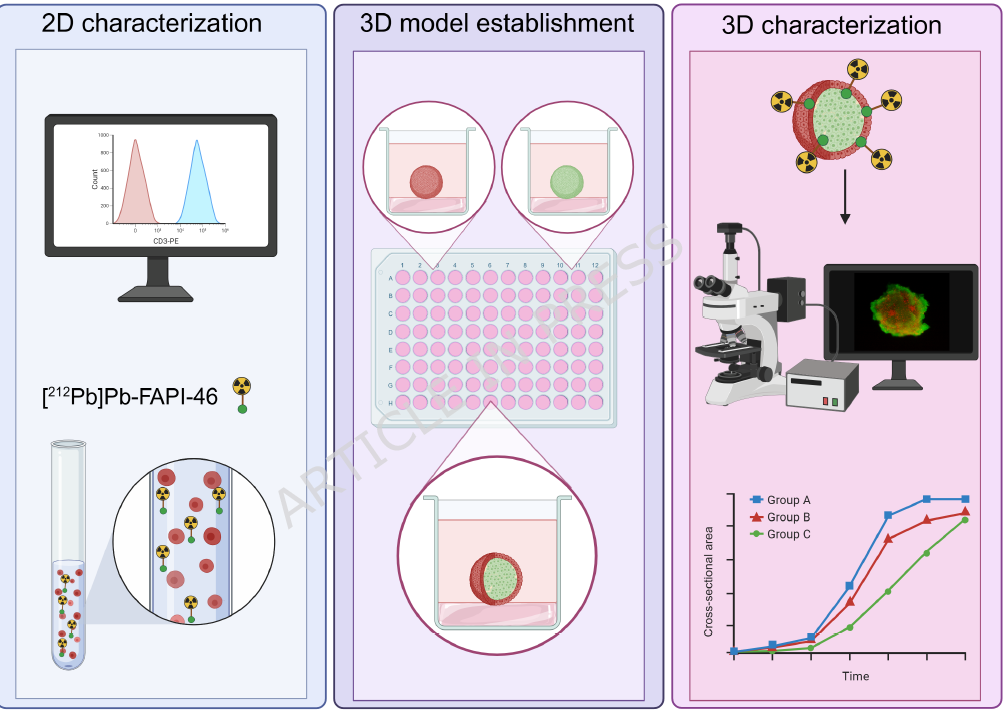
![A 3D co-culture model with fibroblast-restricted FAP expression for cancer therapy using [212Pb]Pb-FAPI-46](https://sofie.com/wp-content/uploads/2026/06/A-3D-co-culture-model-with-fibroblast-500x383.png)