Torsten Kuwert, Christian Schmidkonz, Olaf Prante, Georg Schett, Andreas Ramming
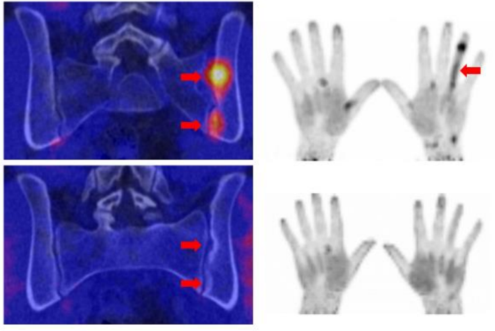
Abstract: In vivo visualization of inflammatory lesions in the body has been revolutionized by positron emission tomography (PET) with F-18-deoxyglucose (FDG) as a tracer and by magnetic resonance imaging (MRI) with gadolinium-labelled contrast media. Apart from other indications, FDG-PET and MRI have substantially improved the diagnosis and monitoring of immune-mediated inflammatory diseases such as arthritis and connective tissue diseases. While the visualization of active inflammation is well established, the detection of tissue response and tissue remodelling processes, which accompany immune-mediated inflammatory diseases (IMIDs) and lead to organ damage, is not well established. Tissue remodelling processes during inflammation are based on mesenchymal stroma cell activation and expansion in parenchymatous organs or the synovial membrane of inflamed joints. These cells express specific markers, such as fibroblast activation protein (FAP), that can be visualized by radiolabelled compounds (e.g. FAP inhibitors; FAPI) using PET. First evidence shows that focal accumulation of FAPI tracer, indicating active tissue remodelling, is observed in patients with IMIDs that are characterized by a combination of chronic inflammation and tissue responses, such as systemic sclerosis, IgG4 syndrome, or spondyloarthritis. Such FAPI-positive remodelling lesions are not always FDG-positive indicating that inflammation and tissue responses can be disentangled by such methods. These data suggest that tracers such as FAPI allow to visualize the dynamics of tissue responses in immune-mediated inflammatory diseases in vivo. This development opens new options for early recognition of tissue remodelling in the context of chronic inflammation.


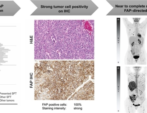
![FAP Expression in Renal Tumors Assessed by [68Ga]Ga-FAPI-46 PET Imaging and FAP Immunohistochemistry: A Case Series of Six Patients](https://sofie.com/wp-content/uploads/2025/12/info.ibamolecular-scaled-500x383.jpg)


![[68Ga]Ga-API-46 PET accuracy for cancer imaging with histopathology validation: a single-centre, single-arm, interventional, phase 2 trial](https://sofie.com/wp-content/uploads/2025/09/image-500x383.png)
