
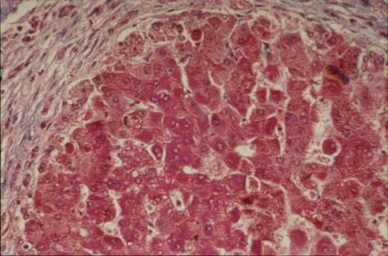
Abstract: Ga-FAPI PET/CT was performed in a 56-year-old cirrhotic patient with multiple liver nodules, which were suspected of hepatocellular carcinoma by contrast-enhanced magnetic resonance but were not visualized by F-FDG PET/CT. Increased Ga-FAPI liver uptake was observed in this cirrhotic patient. However, the nodules had no pathological Ga-FAPI uptake and were clearly visualized due to the low activity compared with surrounding liver parenchyma. The Ga-FAPI PET/CT features, suggestive of benign nodules, have been later confirmed by histopathological examination. This case suggested that Ga-FAPI PET/CT may be useful in the differentiation between benign nodules and hepatocellular carcinoma in the patient with liver cirrhosis.
Affiliations:
- From the Departments of Radiation Oncology.
- Nuclear Medicine and Minnan PET Center, Xiamen Cancer Hospital, The First Affiliated Hospital of Xiamen University, Teaching Hospital of Fujian Medical University, Xiamen, China.
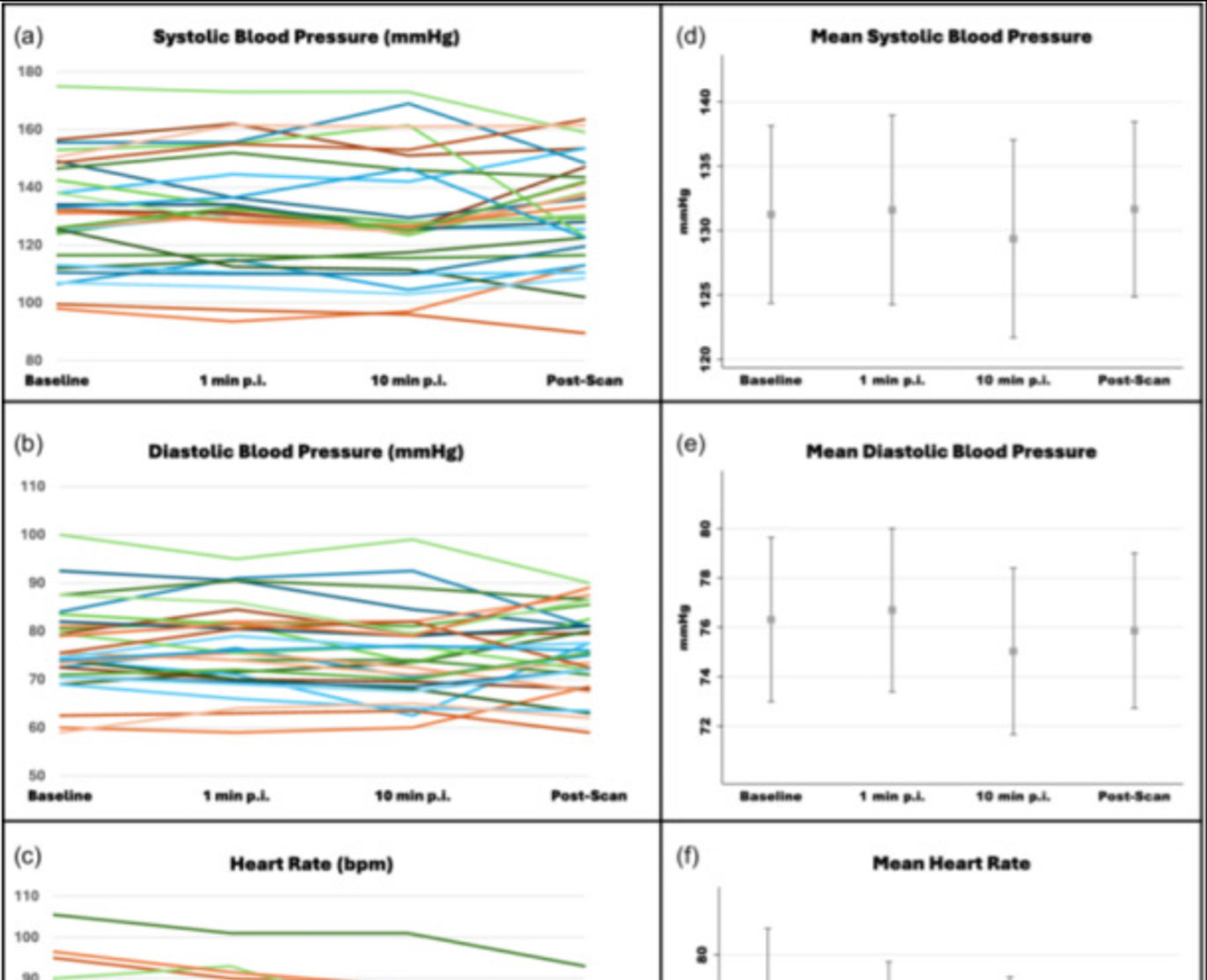
![Safety of [68Ga]Ga-FAPI-46 PET/CT including hemodynamic measurements and patient-reported outcomes in cancer patients](https://sofie.com/wp-content/uploads/2026/05/NewImage-500x383.jpg)
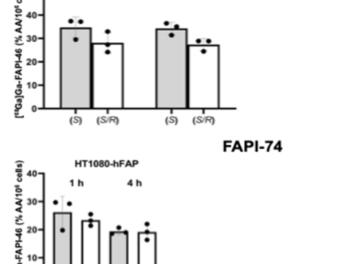
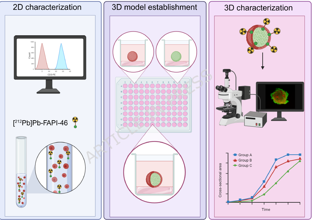
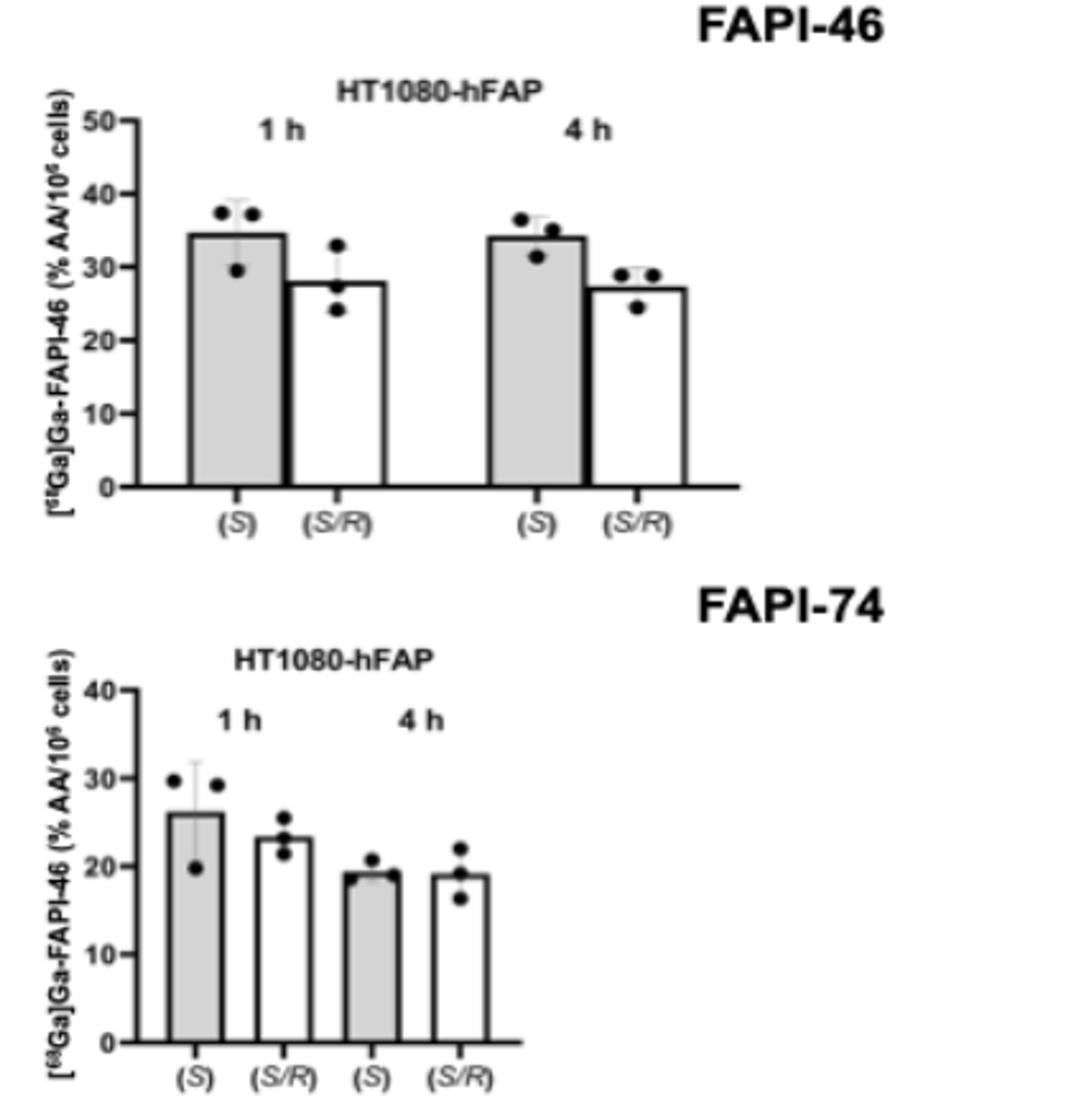
![A 3D co-culture model with fibroblast-restricted FAP expression for cancer therapy using [212Pb]Pb-FAPI-46](https://sofie.com/wp-content/uploads/2026/06/A-3D-co-culture-model-with-fibroblast-500x383.png)