
Sajjad Sadeghpour, Atena Aghaee, Hojjat Ahmadzadehfar, Alessio Rizzo, Giorgio Treglia, Ramin Sadeghi
Abstract:
Background: Regarding the assessment of brain metastatic lesions, [18F]FDG PET/CT encounters challenges due to heightened physiological uptake in normal brain tissue, resulting in poor tumor-to-background ratios (TBR). Detecting brain metastases accurately has long been a challenge with standard imaging methods. The elevated physiological uptake of [18F]FDG in normal brain parenchyma limits its ability to distinguish metastatic lesions owing to poor tumor-to-background contrast. On the other hand, radiolabeled fibroblast activation protein inhibitors (FAPI) have emerged as a newer imaging tracer, showing promise. This study aimed to collect available literature to assess FAPI’s ability to detect brain metastases across various cancer types.
Methods: We reviewed studies published up to April 2025, utilizing different databases, including Google Scholar, PubMed, and Scopus, primarily examining the performance of FAPI based on standardized uptake values (SUVs) and TBRs. Studies focusing on the diagnostic performance of FAPI-based PET for brain metastasis were included. The primary endpoint was the detection rate of FAPI-based tracers.
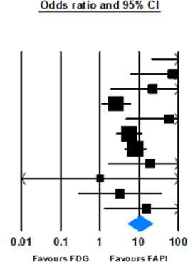
Results: In 24 studies involving over 115 patients and more than 291 lesions, FAPI-based imaging detected of 87.9% lesions (95% CI: 76.1-94.3%, p < 0.001) compared to [18F]FDG by the detection rate of 46.3% (95% CI: 30.6-62.8%, p = 0.667). The comparison of detection rates of these modalities showed an OR of 10.78 (95% CI: 5.15-22.55, p < 0.001). FAPI radiotracers exhibited higher TBR compared to [18F]FDG PET/CT (SMD: 1.51, 95% CI: 0.86-2.16, p < 0.001).
Conclusion: In conclusion, FAPI-based tracers seem to surpass [18F]FDG PET in identifying brain metastases. FAPI imaging can potentially serve as a diagnostic tool that offers benefits over traditional methods. Further research is needed to verify its clinical effectiveness.
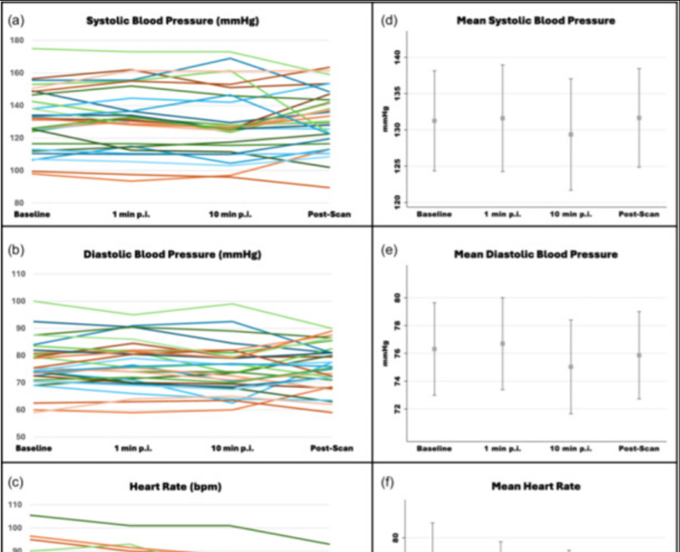
![Safety of [68Ga]Ga-FAPI-46 PET/CT including hemodynamic measurements and patient-reported outcomes in cancer patients](https://sofie.com/wp-content/uploads/2026/05/NewImage-500x383.jpg)
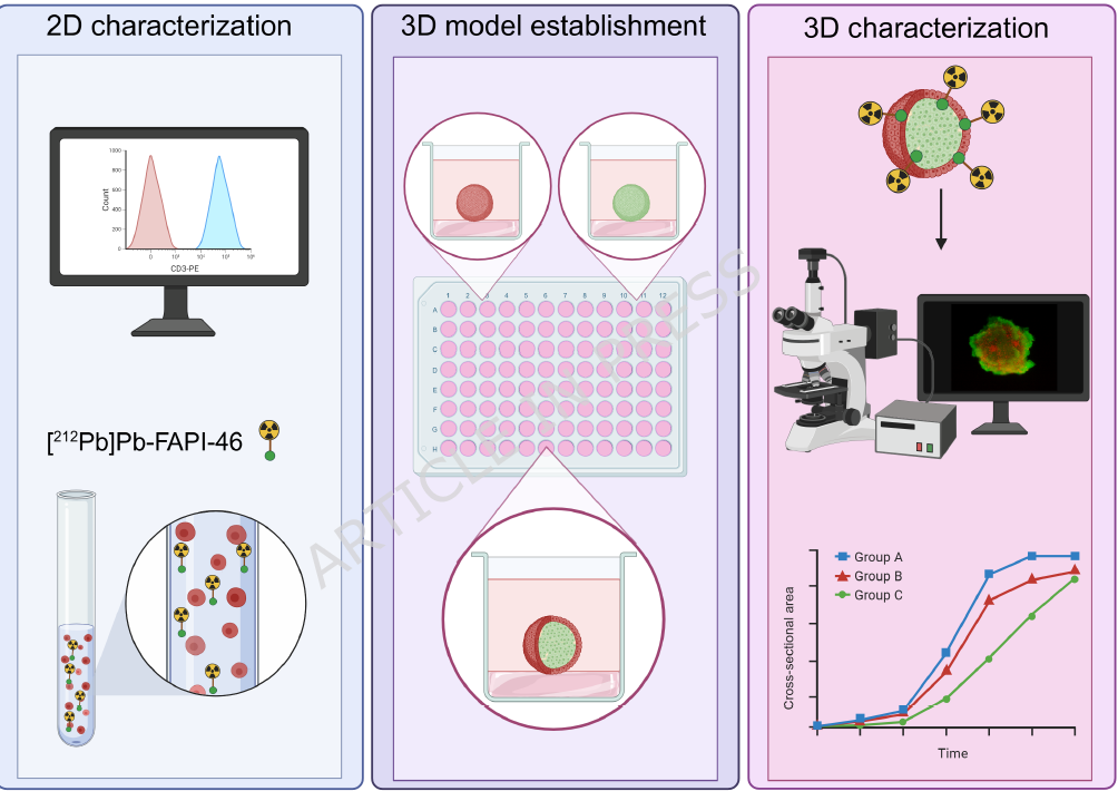
![A 3D co-culture model with fibroblast-restricted FAP expression for cancer therapy using [212Pb]Pb-FAPI-46](https://sofie.com/wp-content/uploads/2026/06/A-3D-co-culture-model-with-fibroblast-500x383.png)