
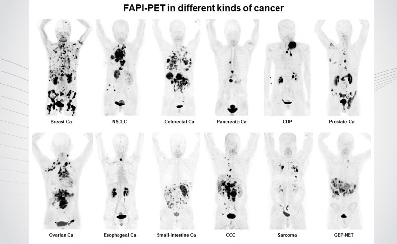
SNMMI 2019 Image of the Year: 68Ga-FAPI-PET/CT in patients reflecting 12 different tumor entities. Ca = cancer; NSCLC = non-small cell lung cancer; CUP = carcinoma of unknown primary; CCC = cholangiocarcinoma; GEP-NET= Gastroenteropancreatic neuroendocrine tumor. Image Credit: Image created with contributions from Clemens Kratochwil, Paul Flechsig, Thomas Lindner, Labidi Abderrahim, Annette Altmann, Walter Mier, Sebastian Adeberg, Hendrik Rathke, Manuel Röhrich, Hauke Winter, Peter Plinkert, Frederik Marme, Matthias Lang, Hans Ulrich Kauczor, Dirk Jaeger, Juergen Debus, Uwe Haberkorn, Frederik L. Giesel; all contributors are affiliated with University Hospital Heidelberg, Germany.
From SNMMI:
A single radiotracer can identify nearly 30 types of cancer, allowing for new applications in noninvasive diagnosis, staging and treatment, according to research presented at the 2019 Annual Meeting of the Society of Nuclear Medicine and Molecular Imaging (SNMMI). The results of the study demonstrate that positron emission tomography/computed tomography (PET/CT) with a fibroblast-activation protein inhibitor (FAPI)—which targets the overexpressed proteins present in cancer—resulted in images with exceptionally clear tumor delineation and high image contrast, as demonstrated in the 68Ga-FAPI-PET/CT image that has been selected as the 2019 SNMMI Image of the Year.
Each year, the SNMMI chooses an image that best exemplifies the most promising advances in the field of nuclear medicine and molecular imaging. The state-of-the-art technologies captured in these images demonstrate the capacity to improve patient care by detecting disease, aiding diagnosis, improving clinical confidence and providing a means of selecting appropriate treatments. This year, the SNMMI Henry N. Wagner, Jr., Image of the Year was chosen from more than 2,300 abstracts submitted to the meeting and voted on by reviewers and the society leadership.
This honor goes to a team of researchers at University Hospital Heidelberg, Germany. Showcasing the efficacy of the FAPI radiotracer, the image demonstrates the uptake of the FAPI in 12 epidemiological tumor entities, including high uptake values in lung, breast, prostate, esophageal and pancreatic cancer. Low background activity was also observed in the various cancers, resulting in high image contrast and excellent tumor delineation. Also, in contrast to FDG-PET/CT, FAPI-PET/CT can be performed without specific patient preparation (fasting, recline during uptake time) after a very short uptake time (~10 min.) and might improve patient comfort and accelerate workflow.
In addition to identifying tumors, the research is also important for the development of cancer treatments in the future. “Immunotherapies can be highly effective in some patients and without any anti-tumor activity in other patients,” noted author Uwe Haberkorn, MD, professor and chair of nuclear medicine at the University Hospital of Heidelberg and the German Cancer Research Center in Heidelberg, Germany. “Currently, predictive biomarkers for appropriate patient selection are limited. Due to its biological role, FAP-targeted diagnostics has the potential to be a predictive biomarker.”
Prof. Haberkorn continued by noting that “The used FAP-ligands contain the DOTA-chelator, which enables labeling with therapeutic radionuclides. The observation that the ligand accumulates in several important tumor-entities potentially indicates a huge field of therapeutic application to be evaluated in the future.”
Furthermore, FAPI-PET already has the potential to add important diagnostic value, particularly in challenging FDG-PET cancer subtypes such as pancreas, ovarian and colorectal cancer and also in patients with peritoneal cancer spread. FAPI-PET might even promote a new route in non-cancer diseased patients such as chronic inflammatory pathogenesis or myocardial infarction, according to Frederik L. Giesel, MD, vice chair of nuclear medicine at the University Hospital Heidelberg.


![SOFIE partners with BAMF Health, diversifying sites for Phase II [68Ga]FAPI-46 study](https://sofie.com/wp-content/uploads/2023/06/Graphic_BAMF_Post-500x383.jpg)